
Our Solution
AI innovation to support sound clinical decisions
UltraSight brings cardiac sonography to more patients in new healthcare settings by simplifying the use of ultrasound technology, allowing more healthcare professionals the ability to screen patients earlier and provide more timely interventions.
The Challenge
Obtaining new skills requires not only time but also training and supervision. Cardiac sonography is a skill that takes years to acquire and requires daily practice to maintain a high level of proficiency.
Today, many healthcare providers lack access to these three key elements: 1) ultrasound devices, 2) mentorship, and 3) the time required to master proficiency in cardiac ultrasound.
At UltraSight we strive to change this paradigm with the power of artificial intelligence. Our goal is to harness the power of AI to close the skillset gap and allow inexperienced users to take cardiac ultrasound images like experts.
Our Solution
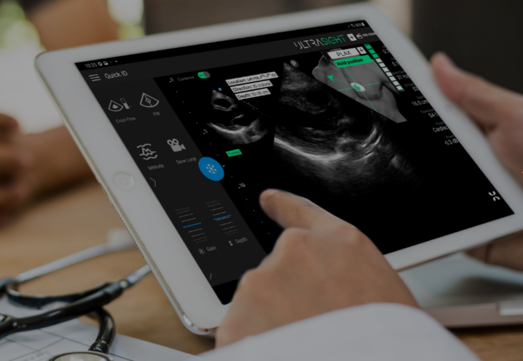
The UltraSight software pairs with PoCUS devices available today and presents guidance instructions as a graphic interface overlaid on top of the ultrasound screen.
Users are guided in real-time how to maneuver the ultrasound probe to obtain a diagnostic quality cardiac ultrasound image.
Once obtained, the image can then be interpreted on the spot or sent to an expert for further evaluation.
How Does
it Work?
UltraSight’s software analyzes the ultrasound stream and guides users through a three-step process of image acquisition:
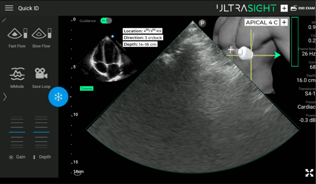
The UltraSight AI algorithm analyzes the cardiac ultrasound image, deducts from the ultrasound image where the ultrasound probe is positioned on patient’s body
Then, the UltraSight software provides real-time instructions how to maneuver the ultrasound probe to move to obtain a diagnostic quality image.
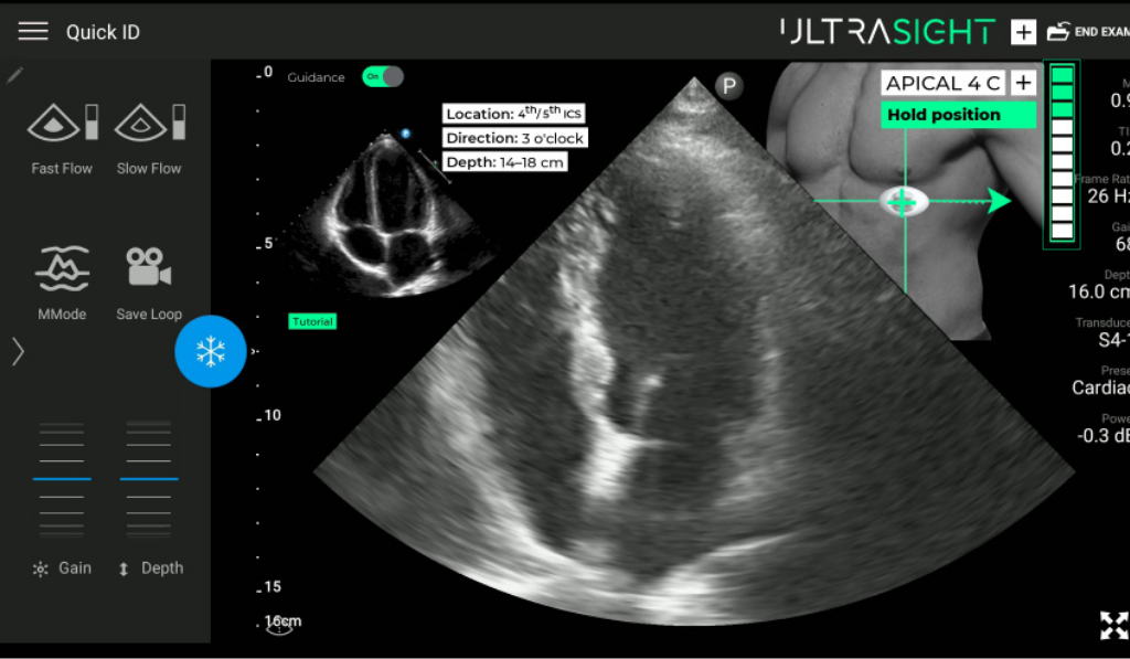
Only when the quality threshold is met, the user is instructed to capture the image
Once the appropriate diagnostic quality image is captured, clinical decisions can be made, or the image can be shared with experts, optimizing clinical decisions.
Unique Features
3D guidance user interface
UltraSight’s Real-Time Guidance software provides instructions on how to move the ultrasound probe in all three dimensions – the X,Y, and Z axes. The instructions are presented using a rich and intuitive graphical user interface.

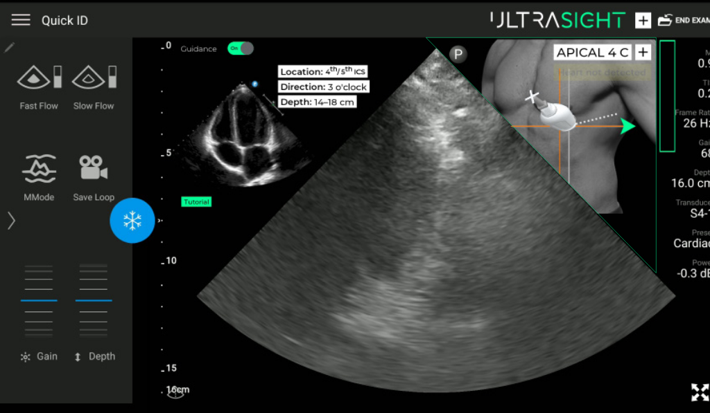
Continuous real-time guidance and movement tracking
UltraSight’s Real-Time Guidance software provides detailed, specific and responsive instructions to the user in real-time. It is both highly sensitive to users’ micro-movements, and seamlessly presents the location of the probe and directions on how to move the probe.

Magnitude
UltraSight’s graphic user interface provides not only the direction in which the user needs to move the probe, but also the precise magnitude of movement. This is done by continuously rendering the relative position of the probe in addition to the ideal location for acquisition.
Clinical Study Validates Technology
UltraSight has successfully concluded its multicenter pivotal study across two U.S. locations, including the University of Chicago and Aurora St. Luke’s Medical Center in Milwaukee, Wisconsin, and at the Sheba Medical Center in Israel. The goal of the study was to evaluate UltraSight AI guidance software’s ability to enable medical professionals without any echocardiography experience to acquire 10 echocardiographic views.
The study results demonstrate that UltraSight Real-Time Guidance can allow medical professionals who do not have prior sonography experience to accurately perform echocardiographic examinations and acquire high quality diagnostic images of the heart in 93-100% of patient exams.
The study also found that the clinical interpretation is similar when exams are done by novice users using UltraSight’s Real-Time Guidance software when compared with sonographers.
Exams With Sufficient Diagnostic Quality To Evaluate These Clinical Parameters:

